


Investigation of the causes of mass fish kills in the Menindee Region NSW over the summer of 2018–2019

This report investigates the causes of three major fish kills in the Darling River near Menindee in December 2018 and January 2019. It was produced at the request of the Hon Bill Shorten MP, Leader of the Federal Opposition and was made public by the Academy at the same time it was provided to Mr Shorten.
Executive summary
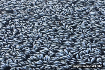
On 15 December 2018 tens of thousands of dead fish were reported along a 30 km stretch of the Darling River near the town of Menindee in New South Wales. High numbers of dead fish were seen in the vicinity of the Old Menindee Weir and Menindee Pump Station. A second, larger fish kill event involving hundreds of thousands of fish was reported on 6 January 2019 on the same stretch of river. A third event followed on 28 January, killing millions of fish. Members of the panel witnessed the beginnings of a fourth event on 4 February 2019.
Many different sectors of Australian society, and of the Menindee region itself, are distressed knowing that fish have been dying en masse, and are concerned about the implications for the health of the river. In addition, these fish are of high cultural significance to Indigenous communities in the region, including those holding Native Title rights.
In response to the first two kills, the Academy was requested by the Leader of the Opposition, the Hon Bill Shorten MP to provide advice on the immediate causes, as well as exacerbating circumstances from water diversions, agricultural runoff or climate change, and to provide recommendations.
Summary findings
The Academy panel made the following findings, illustrated in Figure 1:
- The three fish kills that occurred in rapid succession over December 2018 and January 2019 were unusual in the combination of their severity, impact on large, 20-year-old and older Murray cod, and association with low flows.
- The immediate cause of the fish deaths was stratification and then mixing of a large volume of oxygen-depleted bottom water with the smaller oxygenated surface layer. Conditions such as low- and no-flows and hot temperatures favoured growth of large blue-green algae blooms as well as separation of water layers. As the blooms died and sank they fed bottom layer microorganisms, which used up all available oxygen. Sudden drops in temperature then triggered mixing between the surface and bottom layers, lowering the overall concentration of oxygen in the water beyond the ability to support respiration of the fish. The extreme maximum temperatures, among the hottest on record, are as expected under anthropogenic warming.
- The conditions leading to this event are an interaction between a severe (but not unprecedented) drought and, more significantly, excess upstream diversion of water for irrigation. Prior releases of water from Menindee Lakes contributed to lack of local reserves.
- The root cause of the fish kills is that there is not enough water in the Darling system to avoid catastrophic decline of condition through dry periods. This is despite a substantial body of scientific research that points to the need for appropriate flow regimes. Similarly, engagement with local residents, Indigenous and non-Indigenous, has been cursory at best, resulting in insufficient use of their knowledge and engagement around how the system is best managed.
- The panel strongly supports the objectives of the Water Act 2007 and the framework of the Murray–Darling Basin Plan (2012), which were developed with bipartisan political support and intended to increase water for the environment. However, the findings summarised above and detailed in the following sections point to serious deficiencies in governance and management, which collectively have eroded the intent of the Water Act 2007 and implementation of the Murray–Darling Basin Plan (2012) framework.
The freshwater systems of the Darling are already listed as endangered (NSW, 2007) and include multiple fish species listed as threatened by the Commonwealth. Failure to act resolutely and quickly on the fundamental cause – insufficient flows – threatens the viability of the Darling, the fish, and the communities that depend on it for their livelihoods and wellbeing including the traditional owners, who have recognised rights and responsibilities.
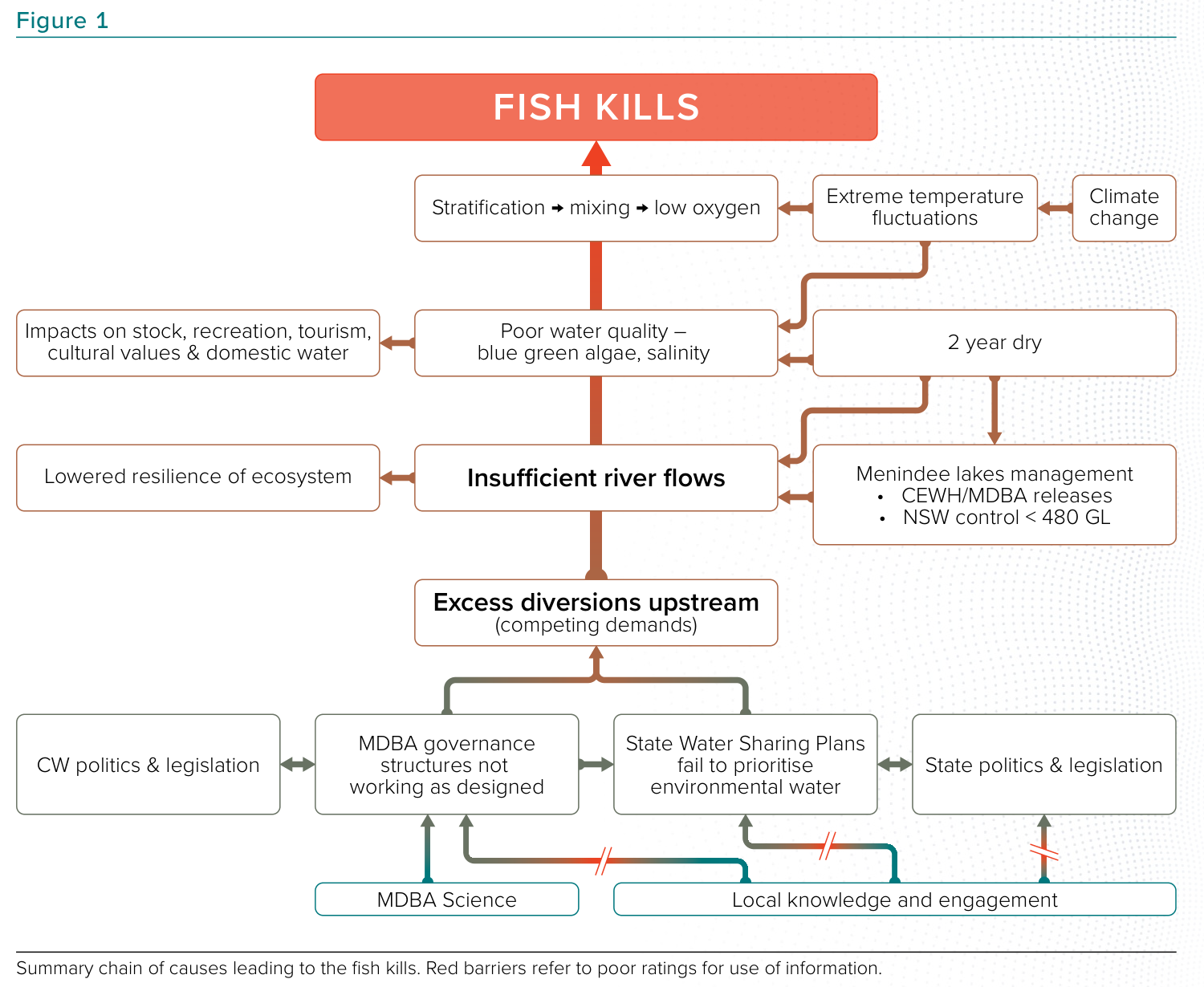
Figure 1: Summary chain of causes leading to the fish kills. Red barriers refer to poor ratings for use of information. MDBA – Murray–Darling Basin Authority; CW – Commonwealth; CEWH – Commonwealth Environmental Water Holder.
Summary recommendations
The Academy expert panel recommends that responsible authorities:
- Within six months, take urgent steps to ensure that there is sufficient flow – considering both quality and quantity of water – in the Darling River to prevent stratification and blue-green algal blooms.
- Within six months, establish a Menindee Lakes restoration project, to determine sustainable management and operation of the lakes system and the Lower Darling and Darling Anabranch.
- Initiate a community planning process in the Lower Darling to restore river health and sustain local livelihoods
- Improve meaningful engagement with river-based communities, including Indigenous peoples.
- Improve the health of the Darling River, through adequate and effective planning, which is scientifically informed.
- Return to the intent of the 2012 Murray–Darling Basin Plan to avoid increasing risks of more fish kills and other environmental problems for the Darling River.
- Invest to fill high priority knowledge gaps as the Murray–Darling Basin Plan continues to be implemented, and then reviewed in 2026.
- Commission within 12 months an independent scientific panel to review progress in implementing the above recommendations.
Expert panel – Terms of reference
The expert panel was asked to provide advice on:
- how the fish kills took place and what caused the magnitude of the event.
- whether water diversions and/or water management practices in the Murray–Darling system have caused or exacerbated the scale of this disaster.
- whether chemical and fertiliser use may have contributed to the event
- what immediate steps can be taken to improve the river system’s health and management within the Basin Plan framework
- whether there has been a step change in inflows due to climate.